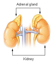
Anatomy of Adrenal gland
The suprarenal or adrenal glands are endocrine glands which help to maintain water and electrolyte balance.
These also prepare the body for any emergency. These are subjected to hyper- or hypofunctioning. Lack of secretion of the cortical part leads to Addison's disease.
Excessive secretion causes retention of salts and fluids.Tumour of adrenal medulla or pheochromocytoma causes persistent severe hypertension.
Subdivisions
The suprarenal glands are a pair of important endocrine glands situated on the posterior abdominal wall over
the upper pole of the kidneys behind the peritoneum. They are made up of two parts.
1 An outer cortex of mesodermal origin, which secretes a number of steroid hormones.
2 An inner medulla of neural crest origin, which is made up of chromaffin cells and secretes adrenaline and noradrenalin or catecholamines.
Location
Each gland lies in the epigastrium, at the upper pole of the kidney, in front of the crus of the diaphragm, opposite the vertebral end of the 11th intercostal space
and the 12th rib.
Size, shape and weight
Each gland measures 50 mm in height, 30 mm in breadth and 10 mm in thickness. It is approximately one-third of the size of kidney at birth and about one-thirtieth of it in adults. It weighs about 5 g, the medulla
forming one-tenth of the gland. Right suprarenal is triangular or pyramidal in shape and the left is semilunar in shape.
Sheaths
The suprarenal glands are immediately surrounded by areolar tissue containing considerable amount of fat.
Outside the fatty sheath, there is the perirenal fascia. Between the two layers lies the suprarenal gland. The two layers are not fused above the suprarenal. The perirenal space is open and is in
continuity with bare area of liver on the right side and with subphrenic extraperitoneal space on the left side.
The gland is separated from the kidney by a septum.
RIGHT SUPRARENAL GLAND
The right suprarenal gland is triangular to pyramidal in shape. It has:
1.An apex
2. A Base
3 Two surfaces-anterior and posterior.
4 Three borders-anterior, medial and lateral.
LEFT SUPRARENAL GLAND
The left gland is semilunar. It has:
1 Two ends-upper (narrow end) and lower (rounded end).
2 Two borders-medial convex and lateral concave.
3 Two surfaces-anterior and posterior.
Structure and Function
Naked eye examination of a cross-section of the suprarenal gland shows an outer part, called the cortex, which forms the main mass of the gland, and a thin inner part, called the medulla, which forms only about
one-tenth of the gland. The two parts are absolutely distinct from each other structurally, functionally and developmentally.
The cortex is composed of three zones.
a. The outer, zona glomerulosa which produces-mineralocorticoids that affect electrolyte and water balance of the body.
b. The middle, zona fasciculata which produces glucocorticoids.
c. The inner, zona reticularis which produces sex hormones.
The medulla is composed of chromaffin cells that secrete adrenaline and noradrenalin.
Arterial supply
Each gland is supplied by:
1 The superior suprarenal artery, a branch of the inferior phrenic artery.
2 The middle suprarenal artery, a branch of the abdominal aorta.
3 The inferior suprarenal artery, a branch of the renal artery.
Venous drainage
Each gland is drained by one vein. The right
suprarenal vein drains into the inferior vena cava, and the left suprarenal vein into the left renal vein.
Lymphatic drainage
Lymphatics from the suprarenal glands drain into the lateral aortic nodes.
The chromaffin cells in it are considered homologous with postganglionic sympathetic neurons.
Nerve supply
The suprarenal medulla has a rich nerve supply through myelinated preganglionic sympathetic fibres.The chromaffin cells in it are considered homologous with postganglionic sympathetic neurons.


Comments
Post a Comment